 Ocularis Review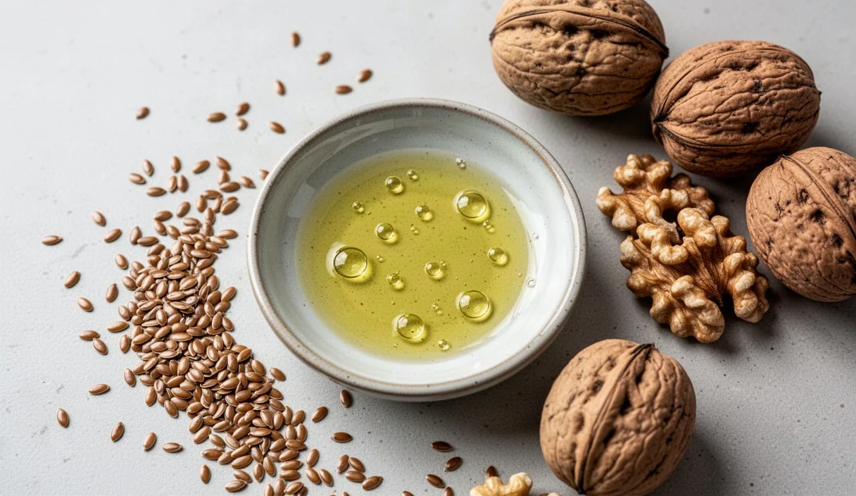
Evaporative dry eye disease represents the most common subclass of ocular surface disorders encountered in clinical optometry and ophthalmology. Unlike aqueous-deficient dry eye, which stems from insufficient fluid production by the lacrimal glands, evaporative dry eye originates primarily from an unstable tear film compromised by meibomian gland dysfunction. When the outermost lipid barrier breaks down prematurely, ambient airflow and thermal evaporation strip moisture from the underlying corneal epithelium, sparking a cascade of hyperosmolarity, friction, and chronic inflammation.
For more than two decades, oral supplementation with long-chain polyunsaturated omega-3 fatty acids has served as a cornerstone of supportive therapy for patients struggling with persistent evaporative symptoms. Clinicians frequently recommend eicosapentaenoic acid (EPA) and docosahexaenoic acid (DHA) to improve the quality of lipid secretions and diminish surface irritation. However, shifting clinical trial data, varied supplement formulations, and individual metabolic responses require a closer look at the actual physiological mechanisms, clinical evidence, and daily practicalities of omega-3 supplementation.
Anatomy of the tear film and meibomian lipid layer
The preocular tear film is a structured fluid dynamic system approximately 3 to 5 micrometers thick. Historically described as a rigid three-layer sandwich, contemporary research models view the tear film as a graded two-phase structure: an inner mucoaqueous layer produced by conjunctival goblet cells and the main and accessory lacrimal glands, topped by an ultra-thin outer lipid layer. The mucoaqueous base provides hydration, oxygenation, and nutrient transport to the avascular corneal epithelium, while specialized mucins anchored to the epithelial microplicae ensure that the aqueous fluid wets the naturally hydrophobic ocular surface evenly.
The overlying lipid layer, measuring roughly 40 to 100 nanometers in thickness, is synthesized and secreted by the meibomian glands. These modified sebaceous glands, numbering between 25 and 40 in the upper tarsal plate and 20 to 30 in the lower tarsal plate, deliver their secretions through terminal orifices situated along the eyelid margins, directly anterior to the mucocutaneous junction. During a complete blink, muscular contraction by the pretarsal orbicularis oculi and Riolan's muscle compresses the gland ducts, expressing meibum onto the tear meniscus, where it spreads upward across the ocular surface to form the protective exterior boundary.
Chemically, healthy meibum is a complex mixture of non-polar lipids (roughly 95 percent of total volume, predominantly wax esters, cholesteryl esters, and small amounts of triglycerides and free fatty acids) and polar lipids (primarily phospholipids and sphingolipids). The polar lipids align themselves at the interface with the aqueous phase, acting as a surfactant that allows the thicker, non-polar hydrophobic wax and sterol esters to spread smoothly over the eye. When meibomian gland secretions become inspissated, thickened, or altered in chemical composition, the lipid layer develops patchy discontinuities. Without a coherent non-polar barrier, the rate of tear film evaporation can accelerate fourfold to tenfold, exposing naked nerve endings on the cornea to physical friction and hyperosmolar stress.
Proposed anti-inflammatory mechanisms of EPA and DHA
The primary therapeutic rationale for systemic supplementation with omega-3 fatty acids centers on shifting the systemic and local balance of lipid mediators. Both omega-6 fatty acids (such as arachidonic acid, or AA) and omega-3 fatty acids (specifically EPA and DHA) compete for the same cyclooxygenase (COX) and lipoxygenase (LOX) enzymatic pathways within cellular membranes. Diets heavily skewed toward refined seed oils deliver an excess of arachidonic acid, yielding pro-inflammatory eicosanoids, including prostaglandin E2 (PGE2), thromboxane A2, and leukotriene B4 (LTB4). These mediators promote leukocyte infiltration, increase vascular permeability in the conjunctiva, and induce inflammatory changes within the meibomian acini.
When systemic intake of EPA and DHA increases, these molecules incorporate directly into the phospholipid bilayers of immune cells, lacrimal acinar cells, and meibomian gland tissue. EPA directly competes with arachidonic acid for COX and LOX enzymes, shifting downstream production toward three-series prostaglandins (such as PGE3) and five-series leukotrienes (such as LTB5). These alternative metabolites exhibit substantially weaker inflammatory potency. For example, LTB5 possesses less than 10 percent of the chemotactic potency of LTB4, thereby curtailing the recruitment of neutrophils and macrophages to the inflamed eyelid margins.
Beyond competitive inhibition, both EPA and DHA act as biochemical precursors for specialized pro-resolving mediators (SPMs). Enzymatic conversion of EPA yields E-series resolvins (RvE1, RvE2), whereas DHA conversion yields D-series resolvins (RvD1 through RvD6), protectins (such as neuroprotectin D1), and maresins. These active chemical signals do not merely suppress immune responses; they actively direct the resolution phase of inflammation by:
- Stopping further neutrophil migration across vascular endothelium into the ocular tissue.
- Stimulating non-phlogistic macrophage phagocytosis of apoptotic cellular debris and oxidized meibomian lipids.
- Downregulating nuclear factor kappa-light-chain-enhancer of activated B cells (NF-kB), dampening the expression of pro-inflammatory cytokines like interleukin-1 beta (IL-1b), interleukin-6 (IL-6), and tumor necrosis factor-alpha (TNF-a).
- Restoring baseline corneal nerve sensitivity and protecting goblet cell densities in the bulbar conjunctiva.
The DREAM trial methodology and controversial findings
The relationship between omega-3 fatty acids and dry eye syndrome was re-evaluated following the publication of the Dry Eye Assessment and Management (DREAM) study in 2018. Funded by the National Eye Institute, this multicenter, double-masked, randomized clinical trial enrolled 535 subjects who displayed moderate-to-severe dry eye symptoms despite prior medical therapy. The active intervention group received a daily oral dose of 3000 milligrams of fish-derived omega-3 fatty acids (composed of roughly 2000 mg EPA and 1000 mg DHA), while the control group received an identical-appearing daily dose of 5000 milligrams of refined olive oil, which provided approximately 3300 mg of oleic acid.
Over the course of 12 months, researchers tracked the Ocular Surface Disease Index (OSDI) score as the primary outcome measure, alongside secondary clinical markers including tear break-up time (TBUT), corneal fluorescein staining, Schirmer's test, and conjunctival staining. Both groups exhibited significant, sustained symptom reduction: mean OSDI scores dropped by 13.9 points in the active omega-3 group and by 12.5 points in the olive oil control group. Because the difference between the two cohorts failed to achieve statistical significance (p = 0.61), investigators concluded that high-dose omega-3 supplementation was not superior to the olive oil placebo for treating dry eye signs and symptoms.
The DREAM trial sparked debate among ocular surface specialists regarding trial design and interpretation. First, the trial permitted participants in both arms to continue using their existing ocular treatments, including topical artificial tears, cyclosporine, warm compresses, and punctal plugs. This baseline flexibility obscured the specific isolated contribution of the fatty acid intervention. Second, critics pointed out that refined olive oil is not a biologically inert placebo; oleic acid possesses mild anti-inflammatory properties, and olive oil contains phenolic compounds that might exert local or systemic benefits. Third, red blood cell membrane fatty acid assays within the trial revealed that while the active group achieved clear biochemical uptake of EPA and DHA, a significant fraction of control participants also experienced shifts in fatty acid profiles, suggesting dietary alterations outside the trial protocol.
Differences between fish oil, krill oil, and algal sources
Patients seeking omega-3 supplementation must choose between several distinct lipid vehicles. The bioavailability, stability, and chemical form vary across source materials, impacting both clinical utility and patient compliance.
| Source Type | Primary Chemical Form | Typical EPA:DHA Ratio | Secondary Active Compounds | Practical Considerations |
|---|---|---|---|---|
| Standard Fish Oil | Ethyl Ester (EE) or Re-esterified Triglyceride (rTG) | 3:2 (approx. 180 mg EPA / 120 mg DHA per gram basic) | Trace Vitamin E added as preservative | Lowest cost; rTG forms show higher absorption than EE forms; fishy aftertaste common |
| Krill Oil (Euphausia superba) | Phospholipid-bound (predominantly phosphatidylcholine) | 2:1 (higher EPA, lower DHA relative to fish) | Astaxanthin (natural carotenoid antioxidant) | Higher relative bioavailability per gram; smaller capsule sizes; higher cost per milligram |
| Algal Oil (Schizochytrium sp.) | Natural Triglyceride (TG) | 0.5:1 to 1:2 (typically DHA-dominant) | Trace mixed tocopherols | Strictly vegan; zero marine toxin accumulation risk; bypasses ocean overharvesting concerns |
The molecular configuration of the fatty acid directly influences intestinal absorption. In raw fish, fatty acids occur as natural triglycerides. During concentration and purification processes, manufacturers commonly transesterify these fats into ethyl esters to concentrate EPA and DHA molecules efficiently. Re-esterified triglyceride (rTG) oils undergo a further enzymatic conversion step to re-attach the free fatty acids to a glycerol backbone. Several comparative trials indicate that rTG formulations yield faster and more complete red blood cell incorporation than ethyl ester products, particularly when consumed without a concurrent high-fat meal.
Krill oil carries its omega-3 content bound to phospholipids. Because phospholipids are amphiphilic (both water- and fat-soluble), they self-assemble into microscopic micelles within the gastric fluid without relying on immediate biliary emulsification. This unique property may permit lower absolute doses to achieve equivalent tissue saturation, though head-to-head clinical dry eye trials directly comparing krill oil to high-dose rTG fish oil remain limited in sample size. Algal oil serves as an alternative for strict vegetarians and individuals allergic to seafood. Modern fermentation yields high-purity algal lipids with high DHA concentrations, though patients may need targeted blends if higher EPA ratios are desired for specific anti-inflammatory pathways.
Dosage considerations and gastrointestinal tolerance
Clinical trials that demonstrate improvements in tear film parameters generally employ daily doses ranging from 1000 mg to 3000 mg of combined EPA and DHA. Lower doses often prove insufficient to alter the lipid composition of meibum or yield clinical shifts in tear stability. When reviewing product labels, patients must look at the actual combined weight of EPA and DHA rather than the total amount of fish oil listed on the front of the bottle; a generic 1200 mg fish oil softgel may contain only 360 mg of total active omega-3s, requiring four to eight capsules daily to meet clinical targets.
Gastrointestinal side effects represent the leading cause of patient discontinuation. Common complaints include fishy eructation (burping), nausea, loose stools, dyspepsia, and mild abdominal cramping. Strategies to improve tolerance include:
- Consuming capsules immediately before or during the largest meal of the day, particularly one containing dietary fats, which stimulates digestive bile release and accelerates transit into the duodenum.
- Freezing the capsules prior to consumption, which delays dissolution of the gelatin shell until the capsule passes past the stomach into the small intestine.
- Utilizing enteric-coated formulations designed to resist stomach acid and dissolve exclusively in the neutral pH of the jejunum.
- Dividing the daily dose into two equal portions taken twelve hours apart rather than taking a large single dose.
Safety parameters demand attention at higher dosage thresholds. The European Food Safety Authority (EFSA) considers supplemental intakes of up to 5000 mg of combined EPA and DHA safe for general adult consumption. However, individuals taking oral anticoagulants (such as warfarin or direct oral anticoagulants) or antiplatelet medications (such as aspirin or clopidogrel) should consult their prescribing physician before initiating doses above 2000 mg daily. While modern studies show that omega-3s rarely cause clinically significant bleeding events on their own, their mild antithrombotic effects warrant collaborative oversight in patients with bleeding disorders or scheduled surgical procedures.
Checklist for identifying tear film instability symptoms
Because evaporative dry eye frequently shares clinical features with allergic conjunctivitis, contact lens intolerance, and blepharitis, identifying distinct tear film instability markers helps guide targeted management. The following diagnostic checklist outlines typical symptoms and signs associated with lipid layer failure:
- Fluctuating visual acuity: Vision sharpens immediately after a complete blink but degrades within three to five seconds of steady fixation, particularly during visual tasks like reading, working at a computer, or driving.
- Paradoxical reflex epiphora: The eyes water excessively in cold, dry, or windy environments. Rapid evaporation of the base tear film triggers an emergency sensory reflex via the ophthalmic division of the trigeminal nerve, flooding the ocular surface with thin, aqueous fluid that fails to coat the eye.
- Burning or foreign body sensation: A gritty, sandy sensation that worsens progressively as the day proceeds, typically reaching peak discomfort during evening hours.
- Symptom flare during low-humidity exposure: Heightened discomfort in air-conditioned offices, heated car interiors, aircraft cabins, or environments with ceiling fans running overhead.
- Eyelid margin hypervascularity: Visible telangiectatic vessels running along the posterior lid margin, often coupled with cloudy, solid, or toothpaste-like meibomian secretions upon diagnostic manual expression.
- Shortened tear break-up time: Under slit-lamp examination with sodium fluorescein dye, the tear film breaks up, developing dry dark spots across the corneal surface, in under 10 seconds (with severe evaporative disease frequently displaying break-up in under 5 seconds).
Common mistakes
The most frequent error in omega-3 supplementation for dry eye is premature discontinuation. Fatty acid incorporation into the meibocyte cell membranes and subsequent turnover into secreted meibum requires twelve to sixteen weeks of steady daily consumption. Expecting symptomatic relief within seven to ten days leads to frustration and unnecessary discontinuation.
A second common mistake is relying on dietary sources of alpha-linolenic acid (ALA), such as flaxseed oil, chia seeds, or walnuts, as direct substitutes for EPA and DHA. The human hepatic conversion of plant-based ALA to EPA occurs at a rate under 8 percent in healthy adults, and conversion to DHA is less than 1 percent. While flaxseed oil provides caloric nutrition, it rarely delivers enough EPA or DHA to alter ocular surface inflammation.
Finally, patients frequently view omega-3 supplements as a standalone cure rather than one component of a broader hygiene regimen. Oral lipids can improve the chemical quality of meibum, but they cannot physically unblock an obstructed gland orifice. Failing to pair oral therapy with physical interventions, such as daily warm compresses, eyelid margin cleansing, and blink retraining, leaves stagnant, oxidized meibum trapped within the tarsal plates.
Next steps for ocular surface recovery
Patients experiencing symptoms of evaporative dry eye should begin by scheduling a comprehensive ocular surface evaluation with an optometrist or ophthalmologist. A professional exam can measure tear break-up dynamics, grade meibomian gland architecture using non-invasive infrared meibography, and rule out conditions like Demodex blepharitis, lagophthalmos, or autoimmune Sjögren's syndrome.
If oral omega-3 supplementation is approved by a primary care physician or eye care provider, start with a high-quality re-esterified triglyceride (rTG) fish oil or high-concentration algal oil delivering between 1500 mg and 2000 mg of combined EPA and DHA daily. Take the supplement alongside meals containing healthy fats to enhance absorption and minimize stomach upset. Maintain this dosage consistently for a full twelve-week trial period, tracking daily comfort levels and visual stability. Combine this nutritional strategy with twice-daily thermal eyelid compresses and conscious blink pauses during digital screen use to promote tear film stability.