 Ocularis Review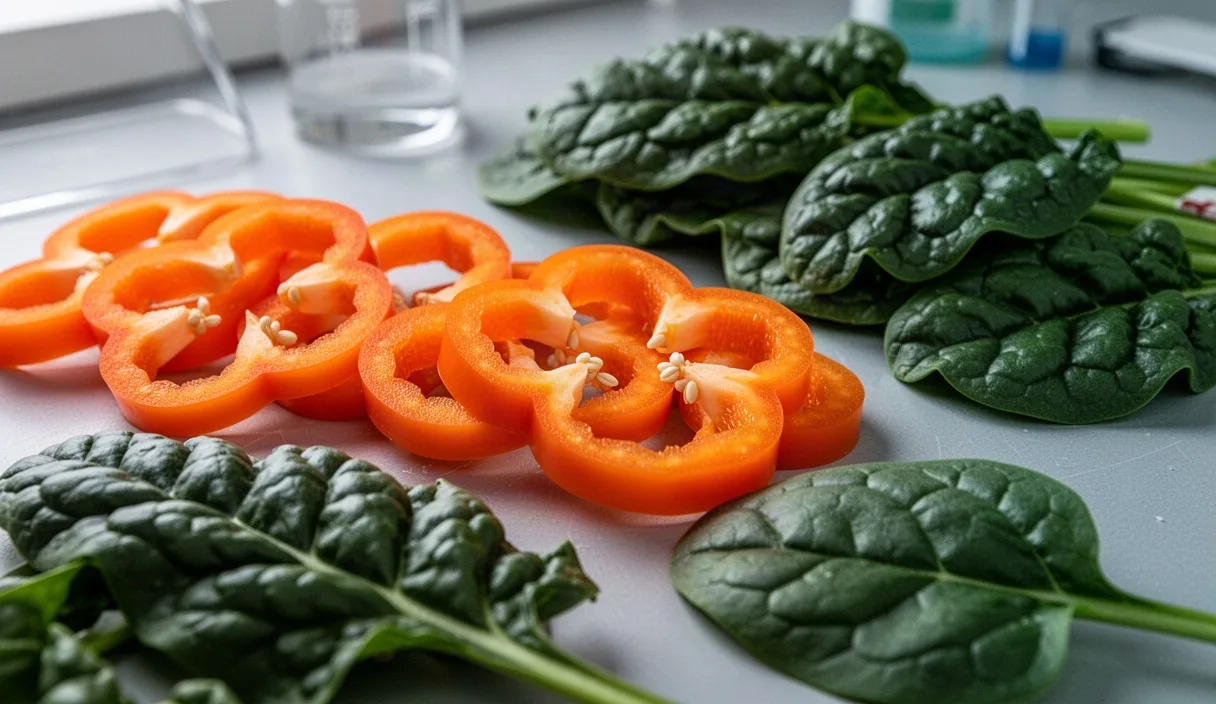
The human visual cycle relies on an uninterrupted supply of vitamin A to synthesize rhodopsin, the light-sensitive photopigment located within the rod photoreceptors of the retina. While preformed vitamin A (retinol) enters the human diet through animal tissues, dairy, and eggs, a substantial portion of the global population secures its retinoid requirements via provitamin A carotenoids found in colored plant matter. Among these pigments, all-trans-beta-carotene holds the highest theoretical yield, possessing a symmetric chemical structure that can yield two molecules of retinol under ideal enzymatic conditions.
The physiological pathway connecting a forkful of cooked carrots or dark leafy greens to the functional retinoid pool within the retinal pigment epithelium (RPE) is intricate. The molecule must first be extracted from its protective vegetable matrix, emulsified into lipid micelles, transported across the mucosal barrier of the enterocyte, split by specific oxygenases, and ferried across the vascular system before crossing the blood-retinal barrier. This biochemical journey is subject to anatomical constraints, dietary composition, and personal genetic variations that alter conversion efficiency.
Basic biochemistry of carotenoid cleavage enzymes
The enzymatic cleavage of beta-carotene into active retinoids occurs primarily through the action of beta-carotene 15,15'-monooxygenase (BCO1), previously designated as BCMO1. This cytosolic enzyme requires non-heme iron (Fe2+) as an essential cofactor and specifically targets the central 15,15' double bond of the symmetric beta-carotene molecule. When oxygen is introduced across this bond, the forty-carbon hydrocarbon backbone splits symmetrically to generate two molecules of all-trans-retinal (retinaldehyde). This process occurs in the mucosal cells of the small intestine, but functional BCO1 expression has also been identified in the liver, kidneys, and ocular tissues, including the retinal pigment epithelium.
Following symmetrical cleavage by BCO1, the resulting all-trans-retinal does not remain in its aldehyde form within the cell. It is rapidly reduced to all-trans-retinol by microsomal enzymes belonging to the short-chain dehydrogenase/reductase family, principally retinol dehydrogenase (RDH) enzymes using NADPH as an electron donor. Once formed, this all-trans-retinol can be esterified into retinyl esters by lecithin:retinol acyltransferase (LRAT) for cellular storage or packaged into chylomicrons for systemic distribution.
A second, eccentric cleavage pathway is mediated by beta-carotene 9',10'-oxygenase (BCO2). Unlike BCO1, BCO2 resides within the inner mitochondrial membrane and carries out asymmetric cleavage at the 9',10' double bond. This reaction does not yield retinal directly; instead, it generates beta-ionone and beta-apo-10'-carotenal. The apo-carotenals undergo subsequent chain shortening through sequential beta-oxidation cycles to yield retinol indirectly, or they are catabolized to prevent carotenoid accumulation. Under standard physiological conditions, central cleavage by BCO1 accounts for the vast majority of systemic vitamin A generation.
| Enzyme | Subcellular Site | Cleavage Position | Primary Reaction Products |
|---|---|---|---|
| BCO1 | Cytosol | Central (15,15' double bond) | Two molecules of all-trans-retinal |
| BCO2 | Mitochondrial matrix/membrane | Eccentric (9',10' double bond) | Beta-apo-10'-carotenal and beta-ionone |
| LRAT | Endoplasmic reticulum | Esterification site | Retinyl esters (storage forms) |
Absorption hurdles in the small intestine
Before any enzymatic cleavage can take place, beta-carotene must clear significant physical hurdles inside the intestinal lumen. Carotenoids are hydrophobic hydrocarbons locked tightly within the crystalline structures or pigment-protein complexes of plant chromoplasts and chloroplasts. Mechanical chewing liberates only a tiny fraction of these pigments. Without thorough breakdown of the cellulose and pectin cell wall matrices, up to 78 percent of ingested beta-carotene remains trapped inside intact plant matter and passes unabsorbed into the colon.
Once released from the dietary matrix, beta-carotene must dissolve within the stomach's lipid droplets before reaching the duodenum. In the upper small intestine, bile acids secreted by the gallbladder and pancreatic lipases disperse these large droplets into mixed micelles. These tiny aggregates, composed of fatty acids, monoglycerides, lysophospholipids, and bile salts, measure roughly 4 to 8 nanometers in diameter. Beta-carotene must partition from the core of dietary oil droplets into these mixed micelles; if it fails to do so, enterocytes cannot take it up.
Apical uptake into the enterocyte is mediated largely by Scavenger Receptor Class B Type I (SR-BI) and Cluster of Determinant 36 (CD36), alongside passive diffusion when luminal concentrations are high. Carotenoids compete with one another for binding sites on these transport proteins. High intakes of lutein or lycopene can reduce the intestinal absorption efficiency of beta-carotene by 20 to 35 percent through competitive inhibition at the SR-BI binding pocket, reducing the net substrate pool available for conversion to retinal.
The necessity of dietary fats for carotenoid uptake
Because beta-carotene is entirely lipophilic, its micellar packaging and subsequent uptake depend on the presence of co-ingested lipids. Without sufficient fat entering the duodenum, the release of cholecystokinin (CCK) remains muted, which prevents robust gallbladder contraction and limits bile secretion. Under these low-fat conditions, mixed micelle formation collapses, and hydrophobic carotenoid molecules aggregate, precipitating out of solution to be excreted in stool.
Clinical feeding trials indicate that a minimum fat threshold is necessary to trigger full mucosal absorption. Diets containing less than 3 grams of fat per meal result in negligible beta-carotene transfer into blood chylomicrons. Increasing lipid content to between 5 and 12 grams per serving produces an exponential rise in area-under-the-curve (AUC) plasma carotenoid concentrations. Fats rich in unsaturated fatty acids, such as oleic acid from cold-pressed oils or linoleic acid from seeds, promote larger micelle surface areas compared to dense saturated animal fats, encouraging higher absorption rates.
- Triglyceride digestion: Pancreatic lipase hydrolyzes triacylglycerols into free fatty acids and 2-monoacylglycerols, the foundational building blocks of mixed micelles.
- Solubilization capacity: Medium-chain triglycerides (MCTs) absorb rapidly into the portal vein but do not form mixed micelles as effectively for long-chain hydrocarbon uptake; long-chain triglycerides (LCTs) remain superior carriers for beta-carotene.
- Biliary stimulation: Co-ingesting 7 grams of olive oil or avocado pulp supplies adequate chemical stimulus to trigger complete gallbladder emptying.
Genetic variations that reduce conversion rates
Conversion rates of beta-carotene to retinol vary widely across individuals. The United States Institute of Medicine established an average dietary Retinol Activity Equivalent (RAE) ratio of 12:1 by weight for beta-carotene from mixed food sources. However, real-world conversion ratios range from 3.8:1 in high converters to more than 28:1 in poor converters. A primary driver of this disparity is genetic polymorphism within the BCO1 gene.
Two common single-nucleotide polymorphisms (SNPs) have a documented impact on enzymatic activity: the Arg267Ser (rs12934922) and Ala379Val (rs7501331) variations. Carrying the Ser267 allele is associated with a 28 percent reduction in BCO1 catalytic activity, while the Val379 variant reduces activity by roughly 21 percent. Individuals who carry both mutations simultaneously exhibit a cumulative reduction of 57 to 69 percent in their ability to cleave beta-carotene into retinal. In these individuals, blood tests reveal high circulating concentrations of parent beta-carotene alongside persistently low levels of retinyl esters following a provitamin A test meal.
Other genetic factors include alterations in the SCARB1 gene, which codes for the SR-BI transporter, and mutations in ISX (Intestine Specific Homeobox). The ISX transcription factor serves as a negative-feedback sensor: when retinoic acid levels within the gut are sufficient, ISX suppresses both SR-BI and BCO1 expression to guard against vitamin A toxicity. Hyperactive ISX variants can prematurely shut down provitamin A absorption even when ocular tissues require additional retinoid delivery.
Retinol delivery to retinal pigment epithelium cells
Once converted into all-trans-retinol inside the gut wall, the molecule is esterified and loaded into chylomicrons. These enter the mesenteric lymphatic system, bypass the liver on first pass, and enter the systemic circulation via the thoracic duct. Peripheral tissues remove a small fraction, but hepatic parenchymal cells clear the balance of the chylomicron remnants. Within the liver, retinyl esters are hydrolyzed back to retinol, transferred to hepatic stellate cells, and stored as stable retinyl palmitate reserves.
When the retina demands functional retinoids, hepatic hydrolases regenerate all-trans-retinol, which binds in a 1:1 stoichiometric ratio to Retinol-Binding Protein 4 (RBP4). This complex enters the blood and binds with transthyretin (TTR), a 55-kilodalton tetrameric transport protein. The formation of the RBP4-TTR complex increases the total molecular weight, preventing the rapid filtration and loss of the small retinol-RBP4 complex through the glomeruli of the kidneys.
The circulating RBP4-TTR-retinol ternary complex travels through the choroidal microvasculature beneath the retina, reaching the basolateral surface of the retinal pigment epithelium. The uptake process into the ocular compartment follows distinct biochemical steps:
- Binding to the membrane receptor: The RBP4-retinol complex docks at STRA6 (Stimulated by Retinoic Acid 6), an integral multi-pass transmembrane pore expressed along the RPE basolateral membrane.
- Translocation across the membrane: STRA6 facilitates the dissociation of retinol from RBP4, driving the hydrophobic molecule across the cell membrane without requiring ATP hydrolysis.
- Intracellular capture: Cellular Retinol-Binding Protein 1 (CRBP1) receives the incoming retinol within the RPE cytoplasm, preventing non-specific lipid membrane intercalation or oxidation.
- Esterification: Lecithin:retinol acyltransferase (LRAT) converts the CRBP1-bound retinol into an insoluble retinyl ester pool.
- Isomerization into the visual pool: The isomerohydrolase RPE65 cleaves and rearranges the all-trans-retinyl ester into 11-cis-retinol.
- Oxidation to visual chromophore: An 11-cis-specific RDH oxidizes 11-cis-retinol to 11-cis-retinal, which is exported across the interphotoreceptor matrix to rods and cones to regenerate light-receptive opsins.
Practical food preparation tips to optimize absorption
Because intact plant matrices form the main physical barrier to provitamin A availability, culinary processing directly dictates how much beta-carotene becomes bioaccessible to intestinal enzymes. Modifying cooking temperatures, mechanical breakdown, and fat pairing can increase carotene extraction fivefold compared to raw consumption.
Thermal softening of plant tissue
Heat weakens cross-linked pectin networks and denatures carotene-associated binding proteins in plant chromoplasts. Steaming or boiling diced carrots, pumpkins, or sweet potatoes for 12 to 18 minutes softens cellular walls without causing thermal degradation of the carotene backbone. Prolonged high-heat frying, by contrast, triggers heat-induced isomerization from the naturally occurring all-trans-beta-carotene to 13-cis or 9-cis isomers, forms that carry 40 to 65 percent less biological activity in conversion pathways.
Mechanical homogenization
Reducing physical particle size increases the exposed surface area for digestive enzymes. Pureeing, blending, or finely grating food breaks a larger proportion of vegetative cells than normal mastication can accomplish alone. Blended spinach or pureed carrot soup exhibits a bioaccessibility rate exceeding 42 percent, whereas consuming whole raw carrot batons yields an average bioaccessibility below 8 percent.
Lipid co-ingestion
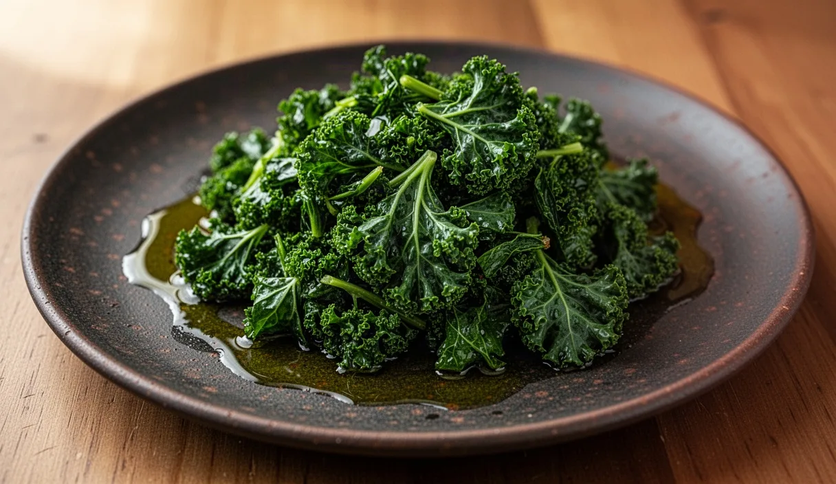
Always prepare carotenoid-dense foods alongside a high-quality lipid source. Adding 6 to 10 grams of culinary lipids (such as extra virgin olive oil, cold-pressed avocado oil, or crushed sesame seeds) to cooked vegetables supplies enough dietary fat to initiate CCK release and support micellar incorporation in the upper small intestine.
Common mistakes
One frequent mistake is consuming large volumes of raw, unprocessed salads or juiced root vegetables without an accompanying lipid source. Green juices composed solely of kale, spinach, and apples provide negligible fat. In this scenario, despite high nominal beta-carotene values on nutrition panels, the intestinal mucosa extracts only trace amounts, shedding the rest as waste.
Another error involves relying entirely on beta-carotene to satisfy retinoid requirements when underlying metabolic conditions reduce conversion. Individuals with hypothyroidism, untreated celiac disease, or chronic pancreatic enzyme insufficiencies struggle to cleave provitamin A efficiently or form mixed micelles. Hypothyroidism downregulates BCO1 transcription, leading to carotenodermia, a benign condition where unconverted yellow carotenes accumulate in the stratum corneum of the palms and soles, while systemic vitamin A status remains marginal.
Finally, using high-dose isolated beta-carotene supplements rather than whole-food sources introduces physiological risks. Synthetic supplements typically flood transport pathways with high concentrations of all-trans monomers, which lack the complex spectrum of co-factors found in whole foods. Clinical research has associated excessive, long-term supplemental beta-carotene with competing transport blocks against other vital ocular pigments, such as lutein and zeaxanthin, which share the same SR-BI entry receptors.
Practical next steps
To establish whether dietary provitamin A meets your biological and ocular needs, consider tracking specific biomarkers alongside meal habits. A registered dietitian or physician can review your diet and run targeted lab tests.
- Request an integrated lipid and retinoid panel: Ask a healthcare professional to assess serum retinol along with parent carotenoid levels (beta-carotene, lutein, lycopene). A high ratio of serum beta-carotene to serum retinol can indicate reduced BCO1 conversion capacity.
- Examine digestive factors: If persistent digestive issues, pale stools, or bloating occur after eating fats, evaluate for bile salt insufficiency, exocrine pancreatic deficiency, or gut absorption disorders that impair micellar assembly.
- Adjust food prep methods: Shift away from raw vegetable consumption for carotene targets. Incorporate gentle heat and mechanical pureeing, and ensure each meal provides at least 5 grams of digestible lipids.
- Personalize your retinoid strategy: If blood testing reveals low serum retinol despite an intake of provitamin A that meets dietary targets, discuss adding preformed retinol (from pasture-raised eggs, oily fish, or dairy) with a qualified clinician.